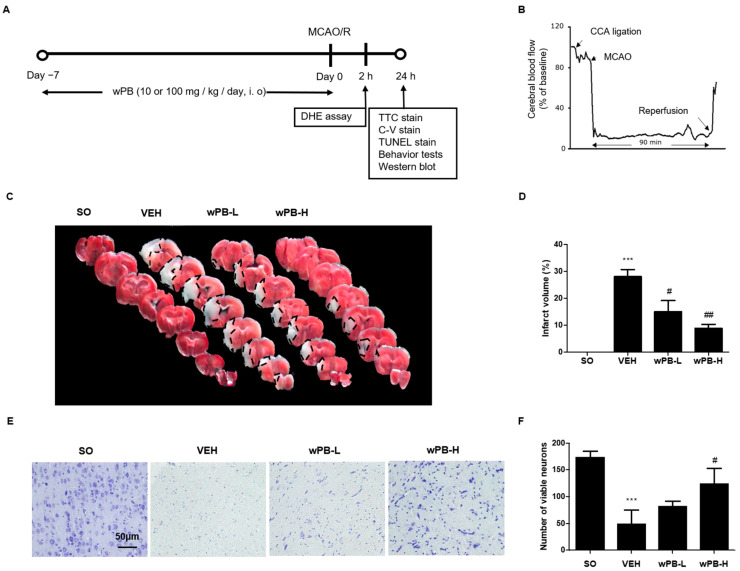
Figure 4.
wPB can minimize infarct volume and neuronal death in mice with focal cerebral ischemia. (A) Timelines of the in vivo experimental protocols and (B) representative Doppler flowmetry showing time-dependent changes in cerebral blood flow values in mice during the MCAO/R operation. (C) Representative photographs showing the TTC-stained brain serial sections and (D) quantitative graphs showing the % area of infarction of different groups. Upon TTC staining, infarcted areas generally appear white. (E) A representative image of C-V-stained ischemic penumbra tissue and (F) quantitative graph showing the average number of viable neurons of the different groups. In all graphs, values are presented as the mean ± standard deviation (*** p < 0.001 vs. the SO group; # p < 0.05; and ## p < 0.01 vs. the VEH group). MCAO/R, middle cerebral artery occlusion and reperfusion; TTC, 2,3,5-Triphenyltetrazolium chloride; C-V, cresyl-violet.

