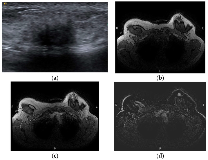
Figure 13.
A woman with a history of left breast cancer presented with a palpable mass 2 years after bilateral mastectomy and implant reconstruction: (a) on US, the mass was hypoechoic with indistinct and irregular margins; (b) on T1-weighted MR images, the mass is hypointense with an irregular shape and spiculated margins; (c) on DCE MRI, 3 min after contrast administration and (d) subtraction image, the mass is strongly and homogeneously enhancing. The diagnosis was fat necrosis after core biopsy.

