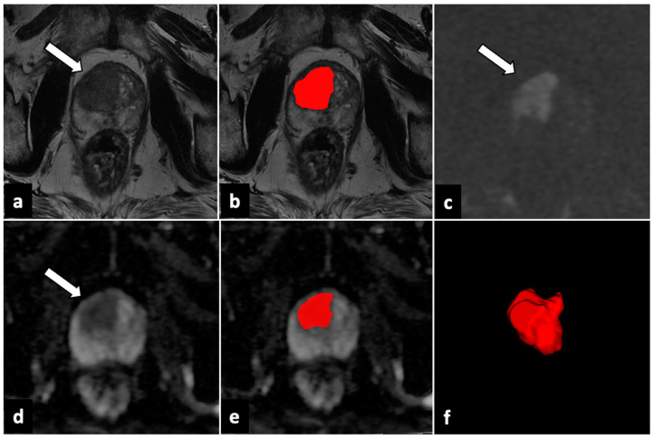
Figure 2.
ROI delineation and generated 3D VOI in a transitional zone lesion. In the right transitional zone of the gland apex, an ill-defined lesion with a major diameter >15 mm is observed (white arrows). The lesion shows a moderately low signal in the T2W sequence (a), a high signal in DWI (c), and signal loss in the ADC map (d), consistent with a PI-RADS 5 category. It was segmented with T2W and ADC (b,e), and a 3D VOI of the lesion (f) was constructed. ADC: apparent diffusion coefficient; DWI: diffusion-weighted imaging; ROI: region of interest; PI-RADS: Prostate Imaging–Reporting and Data System; T2W: T2-weighted; 3D VOI: three-dimensional volume of interest. Red part shows the contouring and final volume obtained from the lesion.

