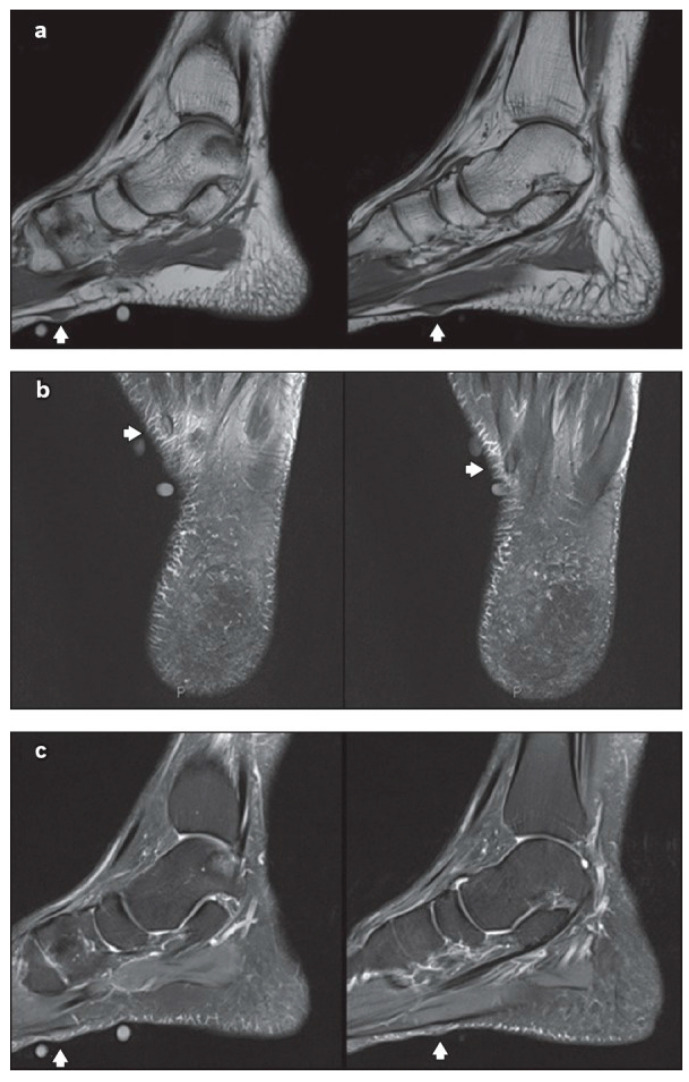
Figure 3.
Non-contrast-enhanced magnetic resonance (MR) imaging showing two sub-centimetre nodules (arrows of a–c) along the inner band of the plantar fascia, on the left foot: (a) sagittal T1-W, (b) axial T2-W fat saturation and (c) sagittal proton density-weighted fat saturation MR images of the left foot. Source: Reprinted from Teo, F.; Mohamed Shah, M.T.; Wong. Clinics in diagnostic imaging. Singapore Med. J. 2019 [37] (Singapore Med. J. licensed under CC BY-NC-SA 4.0, no permission required).

