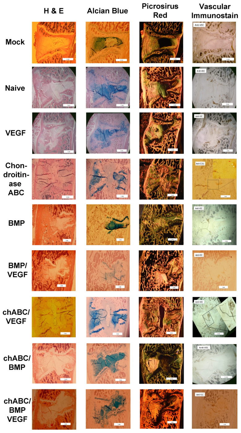
Figure 7.
Representative histology images at 12 weeks after intradiscal delivery of treatments. All images are oriented ventral to the left and cranial as top. Histological staining technique is indicated at the top of each column and treatment group is indicated on the far left of each row. Picrosirus Red birefringence was visualized with circular polarized light, and vascular immunostain shown in the figure is anti-VE-cadherin or anti-CD31 (+), as indicated in each panel in that column (upper-left corner). Arrowheads in immunostained images indicate vascular appearing (red arrowhead) or cell clusters (white arrowheads) with positive immunostain. Scale bars in the right lower corner indicate 1 mm. Chondroitinase ABC is abbreviated as chABC.

