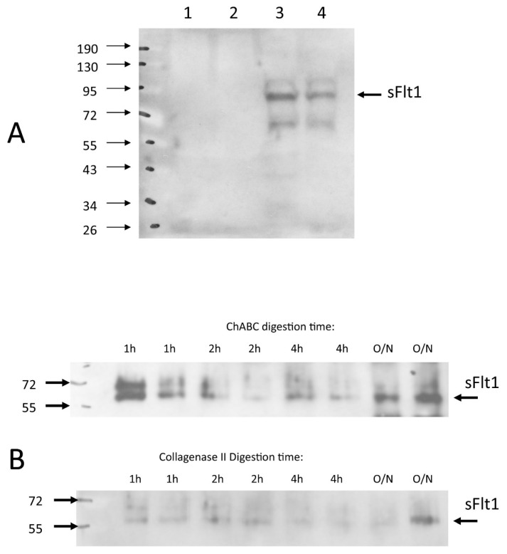
Figure 9.
sFlt Western Blotting of bovine NP cells and rabbit NP tissue. Western immunoblotting for soluble VEGF-R1 (sFlt) demonstrated that sFlt was not detectable in supernatant media in bovine NP monolayers ((A), lanes 1 and 2), but rather was found in the cell/matrix fraction ((A), lanes 3 and 4) and this was not altered by normoxia (21% oxygen culture conditions, lanes 1 and 3) or hypoxia status (2% oxygen culture conditions, lanes 2 and 4). Freshly enucleated rabbit NP tissue when subjected to chondroitinace ABC (ChABC in the figure) digestion over time course (see Materials and Methods) released sFlt from the cell/matrix fraction ((B), upper blot), but digestion of sister samples with Collagenase Type II did not consistently solubilize sFlt ((B), lower blot).

