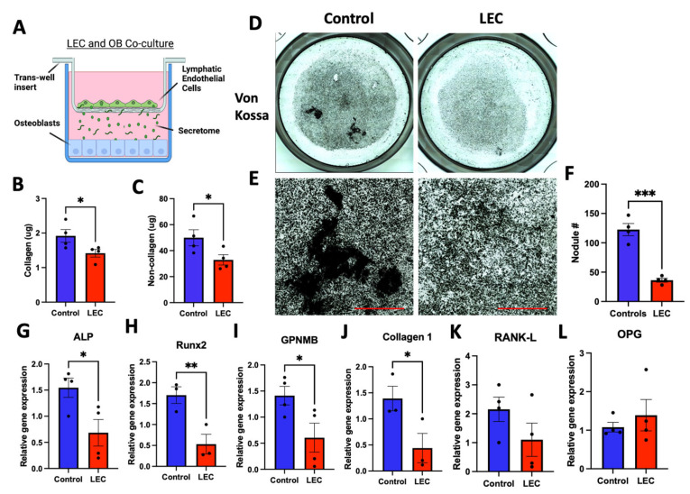
Figure 9.
Co-culture of LECs with differentiating osteoblasts using a trans-well system led to decreased differentiation. MC3T3-E1 cells were cultured with differentiation factors and exposed to the lymphatic secretome from LECs seeded in a trans-well carrier (A). After 1 week of culture, osteoblast matrices were assessed for collagenous (B) and non-collagenous (C) content. After 3 weeks of culture, Von Kossa staining was used to highlight deposits of calcium and potassium (D,E), and this was followed by nodule quantification (F). The gene expression of osteoblast-related markers was assessed after three weeks of culture (G–L). N = 3–4. Data are presented as the mean ± SEM. * p < 0.05, ** p < 0.01, and *** p < 0.001 compared to the untreated control. Scale bar: 1000 μm.

