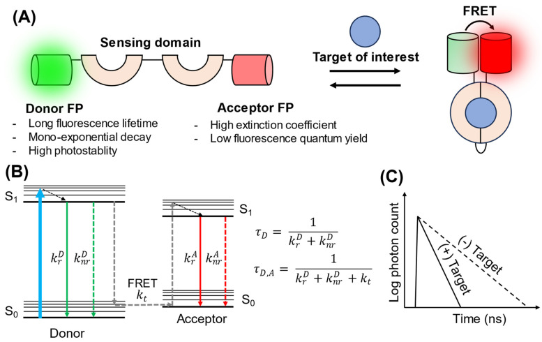
Figure 1.
Schematic illustration of the design and sensing mechanism of FRET–FLIM biosensors. (A) Design of FRET–FLIM biosensors. A donor and acceptor FP are fused to a sensing domain that undergoes a conformational change upon binding to its target. This change brings the two FPs into close proximity, inducing FRET. (B) Jablonski diagram of FRET–FLIM [16]. S0 and S1 represent the ground state and excited states, respectively. Here, is the radiative rate constant of the donor; is the nonradiative rate constant of the donor; is the energy transfer rate constant; is the radiative rate constant of the acceptor; is the nonradiative rate constant of the acceptor; is the fluorescence lifetime when only the donor is present; and is the fluorescence lifetime of the donor in the presence of an acceptor in the FRET pair. (C) Schematic representation of fluorescence decay in the presence and absence of the target. When FRET occurs, elevated kt results in a shortened fluorescence lifetime for the donor.

