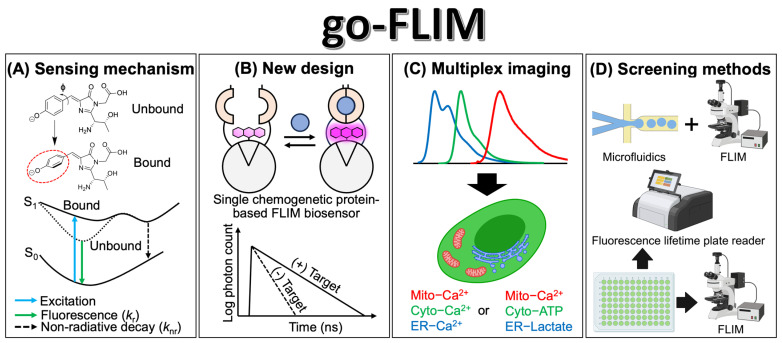
Figure 4.
The promising future of go-FLIM development. (A) A schematic representation illustrating the proposed sensing mechanism of single-FP-based FLIM biosensors. A red circle indicates the rotation of chromophore. (B) Conceptual design of a single chemogenetic protein-based FLIM biosensor and its FLIM response. (C) Representation of multiplex imaging employing go-FLIMs that target various analytes across different organelles such as the mitochondria (mito), cytoplasm (cyto), and endoplasmic reticulum (ER). (D) Introduction of methodologies for the screening of go-FLIM. Illustrations were created with BioRender.com.

