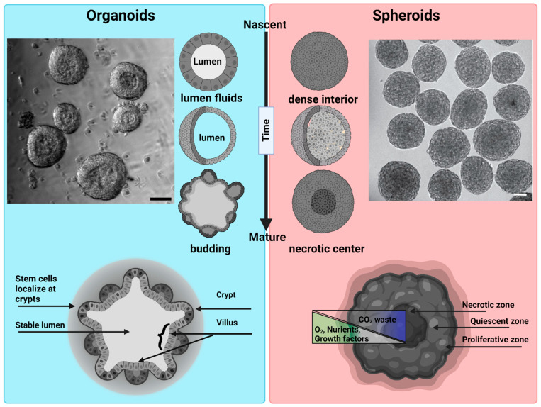
Figure 2.
Morphological differences between organoids and spheroids: Organoids: a representative bright field image of intestinal organoids grown in Matrigel from an intestinal crypt. Nascent organoids have a spherical morphology, are composed of a monolayer of a columnar stem cells and transient amplifying cells, and contain a lumen filled with fluid and extruded cells. As they mature, organoids grow in size and develop a crypt and villus morphology just like intestinal tissue in vivo. The stem cells are localized within the crypts, while the nutrient-absorbing enterocytes form the villus. Intestinal organoids can be used to study mechanisms of barrier function, lumenogenesis, cell division, and cell extrusion. Spheroids: a representative DIC image of spheroids formed by Sarcoma S-180 cells formed via a spinning method within a non-adherent platform. Adopted with permission from [21]. Nascent spheroids are initially roughly spherical, and then merge together to form big spheroids. They are dense structures where cells are connected initially through cell–cell-adhesion. As they mature, they produce necrotic centers. Spheroids are very useful for measuring tumor growth dynamics, mechanical and immuno-resistant barriers, drug responses, and solid stresses. Scale bar is 50 µm.

