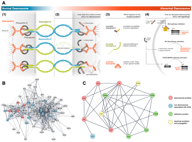
Figure 2.
Architecture of cardiac desmosomes and interactions within the ICD. (A) Schematic of normal and abnormal desmosome architecture, where the abnormal condition reflects reported pathogenic phenotypes in ACM. In a normal desmosome (1), the desmosomal cadherins, desmocollin-2 and desmoglein-2, interact extracellularly to mechanically connect adjacent cells. Intracellularly, plakoglobin and plakophilin-2 serve as adaptor proteins to link the cadherins to desmoplakin. Desmoplakin links the desmosome to the desmin intermediate filament network within the cell. In an abnormal desmosome (2), mutant proteins (3) compromise desmosome structure while causing several other ancillary effects within the cell, including dysregulated cell signaling (4). Hyperactive or inhibitory signaling through plakoglobin or plakophilin-2 can result in a range of ACM-related phenotypes, such as adipogenesis through Wnt inhibition [51], fibrosis through TGFB1 upregulation [52], or recruitment of inflammatory cells to the heart through NF-KB [53]. (B) Graph of interactions between relevant components of the ICD, which include desmosomal proteins (red), non-desmosomal proteins (blue) that have been associated with ACM. Interaction data sourced from STRING database and cross-validated with NCBI gene repositories and various review papers [54,55]. (C) Selected interactive data showing the interconnected links among the desmosomal proteins, AJs, and non-desmosome proteins associated with ACM.

