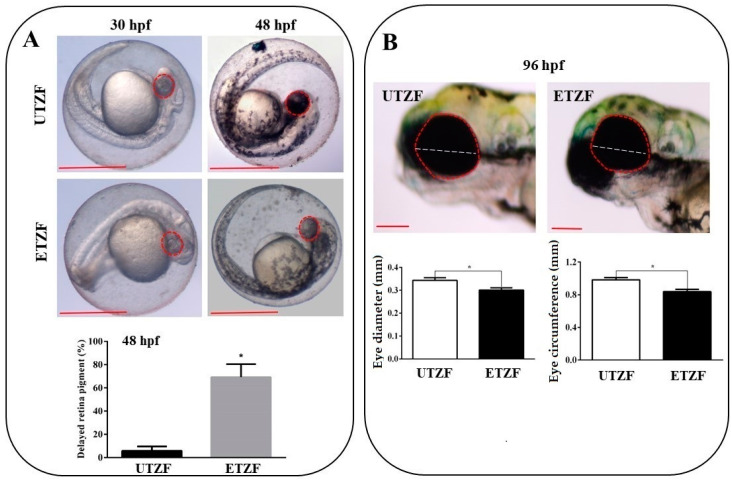
Figure 1.
Etridiazole exposure induced ocular toxicity in zebrafish. (A) Pictures showing the retinal pigment accumulation at 30 and 48 hpf in untreated (upper panel) and etridiazole-treated (lower panel) zebrafish embryos. Graph showing the percentage of eyes with delayed retina pigmentation. (B) Pictures (upper panel) showing eye circumference (red dotted line) and diameter (white dotted line) of untreated and etridiazole-treated zebrafish embryos at 96 hpf. Graphs (lower panels) representing the eye diameter (mm; left graph) and eye circumference (mm; right graph) of the untreated and etridiazole-treated zebrafish embryos. Red dotted circles, eyes; white dotted line, eye diameter; UTZF, untreated zebrafish; ETZF, etridiazole-treated zebrafish. Scale = 0.5 mm. * Statistically significant (p < 0.05).

