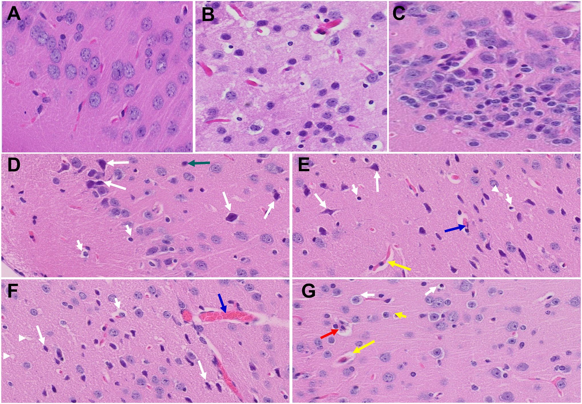
Figure 5.

Acute and long-term changes in brain post-MHV-1 coronavirus infection. (A) Normal mouse brain cortex. (B) Representative image from MHV-1-infected mouse brain cortex showed “perivascular cavitation, congested blood vessel, pericellular halos, darkly stained nuclei, vacuolation of neuropil, pyknotic nuclei, and acute eosinophilic necrosis at 7 days (acute phase)” (Paidas et al., 2021). (C) MHV-1-infected mouse brain cortex (12 months post-infection). (D- G) Enlarged images of (C) showed widespread neuronal necrosis (long arrows), pyknotic nuclei/neuronal clearing (short arrows), vacuolation of neuropil (arrowhead), congested blood vessels (blue arrows), perivascular cavitation (yellow arrows, Virchow–robin space), darkly stained nuclei (green arrow), neuronophagia (red arrow, presence of necrotic neurons surrounded by invaded hypertrophic microglia (G)). (H&E, original magnification 400 × (A-C), and (D-G) are enlarged images of (C))
