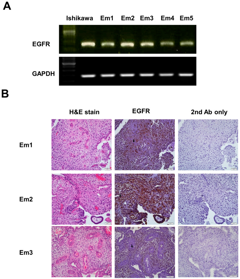
Figure 3.
EGFR protein and mRNA expression in human decidual endometrial tissue and stromal cells. (A) EGFR mRNA expression in human decidual endometrial stromal cells with RT-PCR (Em 1, Em 2, Em 3, Em 4, and Em 5). Ishikawa endometrial cancer cells acted as a positive control. GAPDH was multiplied to verify the same loading. The data are representative of three individual experiments. (B) Immunohistochemical analysis of EGFR protein expression. Brown staining is presented in the middle of three columns describing decidual endometrium sections, including stromal cells (arrow). Sections were counterstained with hematoxylin to demonstrate the nuclei representing decidual endometrium sections in the right of three columns. Sections stained without EGFR antibody serve as a negative control in the left of three columns describing decidual endometrium sections. Micrographs were performed with a 40× objective lens. Scale bars represent 20 μm.

