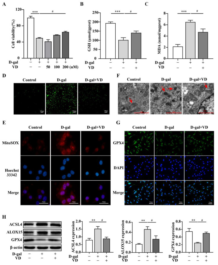
Figure 3.
VD alleviates D-gal-induced ferroptosis in HT22 cells. The HT22 cells were treated with D-gal (250 mM) and VD (0–200 nM) for 24 h. (A) The CCK-8 method was used to detect cell viability (n = 3). (B,C) Intracellular GSH and MDA levels (n = 4). (D) DCFH-DA measured intracellular ROS generation. ROS was green in DCFH-DA fluorescent staining (bar = 100 μm). (E) Mitochondrial ROS levels determined via MitoSOX staining; mitochondrial ROS was red in MitoSOX staining (bar = 50 μm). (F) Transmission electron microscope observation of mitochondrial morphology. The red arrow indicates the mitochondria (bar = 2 μm). (G) Immunofluorescence to detect the expression of GPX4 (bar = 50 μm). (H) Protein expression of ACSL4, ALOX15, and GPX4 (n = 4). Image J software was used to quantify the relative density of ACSL4, ALOX15, and GPX4 relative to β-actin. Compared with the control group, ** p < 0.01, *** p < 0.001; compared with the D-gal group, # p < 0.05.

