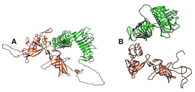
Figure 2.
Ribbon representation of tail fiber proteins 3D structures encoded by the BK025033_ct6IQ4 and phAss-1 genomes. (A) Specified regions of the tail fiber protein of BK025033_ct6IQ4 are marked with red (1–475 aa, two structural domains located at the N-terminal part of the protein) and green (476–1069 aa, two pectate lyase domains located at the C-terminal part of the protein). (B) Structural tail proteins of phAss-1 that correspond to orthologous regions of the tail fiber protein of BK025033_ct6IQ4 are marked with the same colors—red for the tail fiber protein and green for the protein containing two pectate lyase domains. 3D models were predicted using AlphaFold2 and rendered using UCSF Chimera molecular visualizer, version 1.17.

