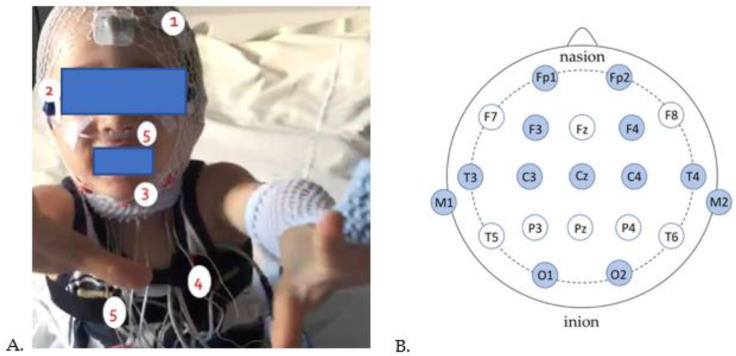
Figure 1.
Pediatric subject fitted for polysomnography recording. (A) 1: Electroencephalogram (EEG), 2: Electrooculogram (EOG), 3: Electromyogram (EMG), 4: Electrocardiogram (ECG), 5: nasal pressure cannula and chest straps. (B) Schematic representation of EEG electrode placement on the scalp (in dark blue are the electrodes intended for our protocol). From front to back, the electrode letter labeling is as follows: Fp (pre-frontal or frontal pole), F (frontal), C (central line of the brain), T (temporal), P (parietal), and O (occipital).

