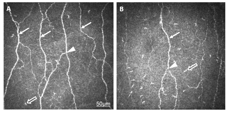
Figure 1.
Representative in vivo confocal microscopic images of the corneal subepithelial nerves in a healthy eye (A) and the eye of a patient with AS (B). The lower density (CNFD) and reduced thickness and length (CNFL) of subepithelial nerve plexi (arrow) in AS compared to normal eyes are demonstrated. The number of branches (CNBD, CTBD, arrowhead) has decreased in AS. An increased number of Langerhans cells (empty arrow) is shown in the level of nerve plexi. The bar represents 50 µm.

