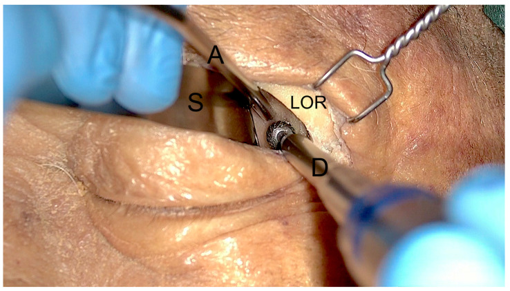
Figure 2.
Exoscopic visualization during the first steps of the surgical approach. After superior eyelid incision and subcoutaneous dissection, the lateral orbital rim (LOR) is identified, and a malleable spatula (S) is used to displace medially the orbital content. Two instruments, a low-profile high-speed drill (D) and an aspirator (A), can be inserted inside the surgical corridor along the lateral orbital wall.

