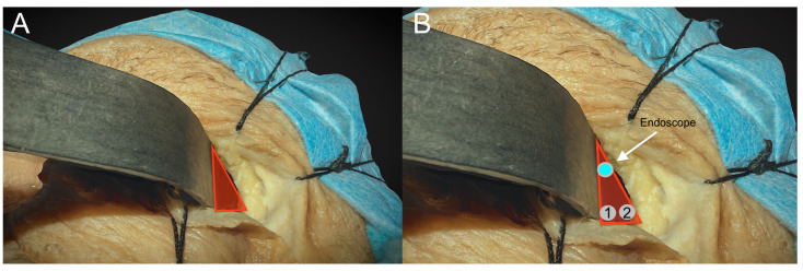
Figure 5.
The picture shows the anatomical boundaries of the triangular port. (A). The lateral margin is represented by the lateral orbital rim, the medial margin by the retractor itself, and the base is the lateral aspect of the upper eyelid crease. (B). The places of insertion of the endoscopic tip are on top, and the two additional instruments are below.

