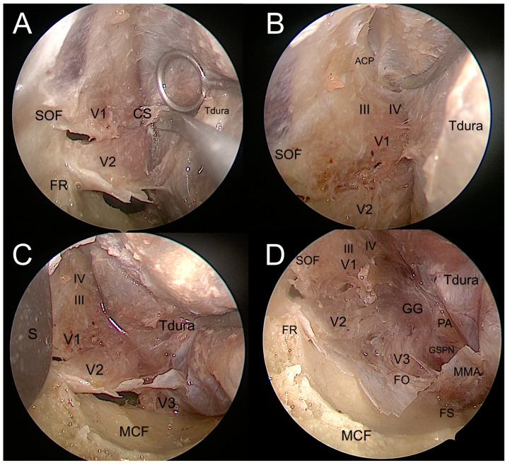
Figure 8.
Interdural dissection of the cavernous sinus. (A). Exposure of the lateral wall of the cavernous sinus. (B). Exposure of the anterior clinoidal process in the upper portion of the surgical field. (C). Exposure of the maxillary and mandibulary nerves in the inferior portion of the surgical field. (D). Exposure of the entire lateral wall of the cavernous sinus, up to the gasserian ganglion and lateral portion of the middle cranial fossa. ACP: anterior clinoidal process; CS: cavernous sinus; FO: foramen ovale; FR: foramen rotundum; FS: foramen spinosum; III: oculomotor nerve; IV: troclear nerve; GG: gasserian ganglion; GSPN: greater superficial petrosal nerve; MCF: middle cranial fossa; MMA: middle meningeal artery; S: spatula; PA: petrous apex; SOF: superior orbital fissure; Tdura: temporal dura; V1: ophthalmic nerve; V2: maxillary nerve; V3: mandibulary nerve.

