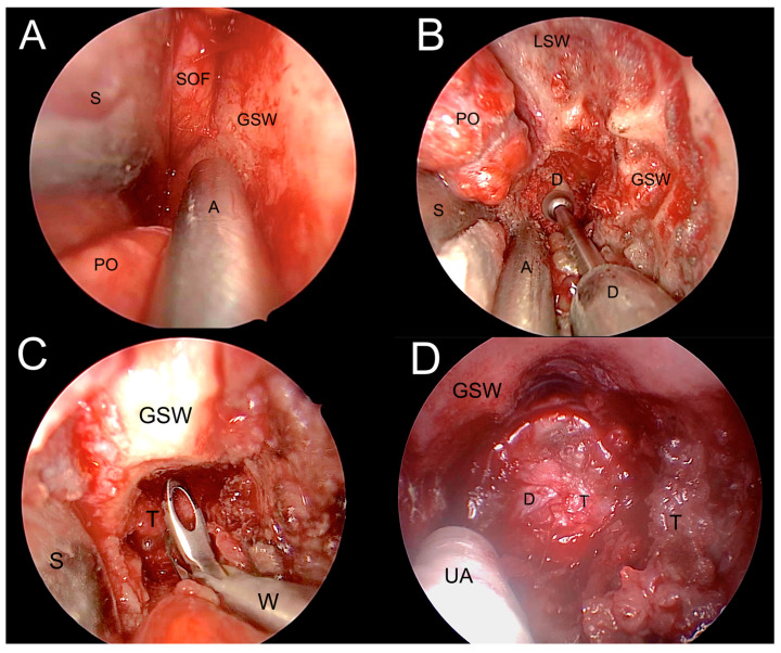
Figure 10.
Transorbital corridor and tumor resection. (A). Exposure of the lateral orbital wall. (B). Drilling the greater sphenoid wing (GSW) to access the middle cranial fossa. (C). Tumor biopsy. (D). Tumor debulking. A: aspirator; D: dura meter; GSW: greater sphenoid wing; LSW: lesser sphenoid wing; PO: periorbita; T: tumor; S: spatula; UA: ultrasonic aspirator; W: Weil nasal forceps.

