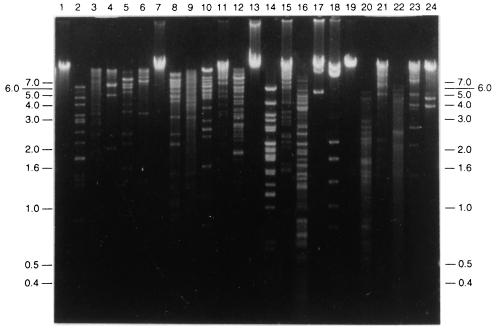
FIG. 3.
Phage DNAs digested with restriction endonucleases to determine molecular weights. Lane 1, uncut T-φD0 DNA; lanes 2 through 6, T-φD0 DNA cut with HpaI, HindIII, BglII, AccI, and EcoRV, respectively; lane 7, uncut T-φD1B DNA; lanes 8 through 12, T-φD1B DNA cut with HapI, AccI, BglII, EcoRI, and EcoRV, respectively; lane 13, uncut T-φD2S DNA; lanes 14 through 18, T-φD2S DNA cut with HpaI, HindIII, AccI, EcoRI, and EcoRV, respectively; lane 19, uncut T-φHSIC DNA; lanes 20 through 24, T-φHSIC cut with HpaI, HindIII, AccI, EcoRI and EcoRV, respectively. The positions of molecular weight standards (in kilobases) are indicated on both sides of the gel.

