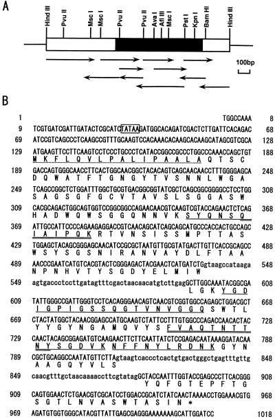
FIG. 3.
Restriction map and sequencing strategy of the genomic DNA for T. reesei egl3 (A) and the complete nucleotide sequence of the gene and deduced amino acid sequence of the EG III protein (B). (A) The HindIII fragment of the egl3 genomic clones is shown as a bar, and the egl3 structural gene region is shown as a filled box. The orientations and lengths of coverage of sequencing primers are shown as horizontal arrows. (B) Intron sequences are in lowercase type. The standard one-letter amino acid code is used. The presumed signal sequence is indicated by the dotted underline. The internal amino acid sequences determined for the lysylendopeptidase-digested peptides of the purified T. reesei EG III are underlined. The amino acid sequences for the design of PCR primers are double underlined.

