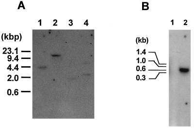
FIG. 5.
Southern hybridization analysis of T. reesei genomic DNA (A) and Northern hybridization analysis of T. reesei RNA (B). (A) Aliquots (20 μg) of T. reesei genomic DNA were digested with each of the following restriction enzymes: BamHI, EcoRI, HindIII, and PstI (lanes 1 to 4, respectively). The resulting fragments were fractionated by agarose gel electrophoresis and then transferred to a nylon membrane for hybridization. The probe used was the egl3 cDNA. The fragments of lambda DNA digested with HindIII were used as molecular size markers. (B) Total RNA samples (10 μg each) were isolated from cells grown in medium containing glucose (lane 1) and Avicel (lane 2). The positions of migration of RNA molecular standards are shown on the left.

