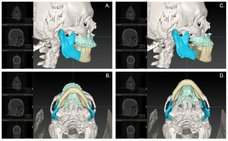
Figure 1.
An example of a virtual plan of an MMA case. Lateral (A) and caudal (B) view of the preoperative 3D virtual hard-tissue skull model of the patient in IPS (KLS Martin, Tuttlingen, Germany). Lateral (C) and caudal (D) view of the postoperative 3D virtual hard-tissue skull model, where the maxilla and mandible are virtually osteotomized according to a Le Fort I osteotomy and BSSO. The maxilla and mandible are advanced and counter-clockwise pitched.

