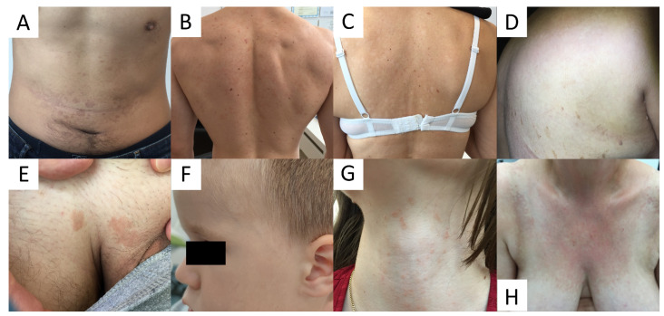
Figure 1.
Variability of clinical presentations of pityriasis versicolor (PV). (A) Hyperpigmented confluent roundish erythemo-desquamative PV macules over the abdomen in a young man. (B) Discrete, achromic confluent PV macules affecting the shoulders and interscapular areas in a young man. (C) Depigmented PV spots on the back in middle-aged women. (D) Discrete, confetti-like PV depigmented spots on the back of an elderly woman. (E) Isolated hyperpigmented “tinea InVersicolor” on the pubis in a young man. (F) Roundish, depigmented macules on the face of a young boy. (G) Minute reddish scaly spots over the neck in a young female. (H) Confluent red scaly macules covering the neckline and presternal area, imitating confluent and reticulated papillomatosis.

