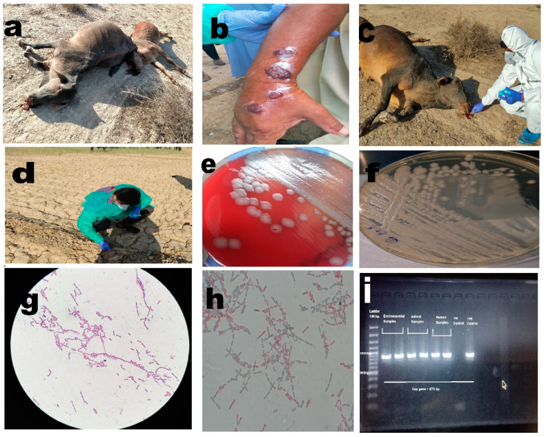
Figure 2.
(a) Anthrax outbreak in animals, illustrating cases of infection among animals. (b) Cutaneous anthrax case in a human, depicting an individual affected by cutaneous anthrax. (c) Collection of soil samples around the carcass, highlighting the process of collecting soil samples in the vicinity of animal carcasses. (d) Collection of plant root samples from infected sites, showing the collection of plant root samples from areas affected by anthrax. (e) Non-hemolytic growth of Bacillus anthracis on blood agar, displaying the characteristic non-hemolytic growth pattern of Bacillus anthracis on blood agar medium. (f) Growth of Bacillus anthracis on PLET agar, illustrating the growth of Bacillus anthracis on PLET agar medium. (g) Bacillus anthracis showing Gram-positive rods in long chains, visualizing the Gram-positive rod-shaped morphology of Bacillus anthracis arranged in elongated chains. (h) Bacillus anthracis with spores, depicting the presence of spores in Bacillus anthracis. (i) PCR bands for Bacillus anthracis capsular gene, showcasing the PCR results demonstrating specific bands corresponding to the capsular gene of Bacillus anthracis.

