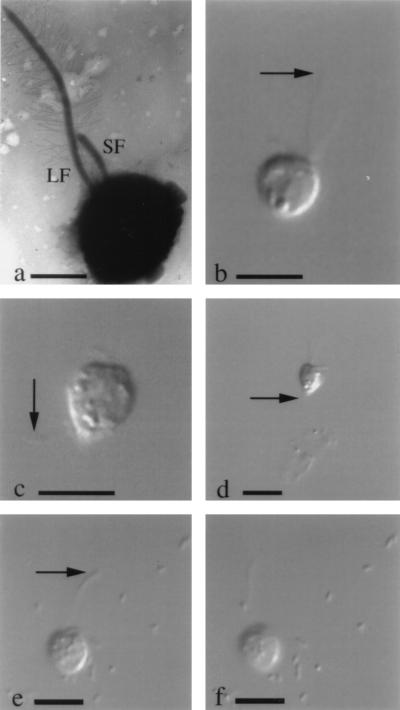
FIG. 2.
Cells of S. guttula from liquid cultures from the MMR aquifer. Micrographs b to f are still images of live cells extracted from digital video clips. (a) Transmission electron micrograph of whole-mount shadow-cast preparation, showing the long flagellum (LF) with flagellar hairs and the short naked flagellum (SF). Scale bar, 2.5 μm. (b) Cell swimming to the rapid beat of the long flagellum; scale bar, 10 μm. (c) Cell which has just become detached from sediment particles (not visible); attachment was achieved by means of a thin posterior protoplasmic filament (arrow). Scale bar, 10 μm. (d) Cell attached to sediment particles. The attachment filament (not visible) arises from the pointed cell posterior (arrow). Scale bar, 10 μm. (e and f) Sequential frames of cell with actively beating long flagellum (arrow). Numerous food bacteria are visible in the background. Scale bar, 10 μm.

