Fig. 4.
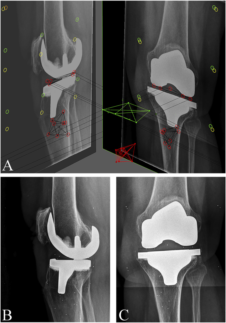
Figs. 4-A, 4-B, and 4-C RSA images of a cemented TKA implant. Fig. 4-A Biplanar (lateral and anteroposterior) views with markers inserted in the polyethylene insert and tibial bone. Fig. 4-B Lateral radiograph of the same implant, which was classified as continuously migrating. Fig. 4-C Anteroposterior radiograph of the same implant.

