Figure 1.
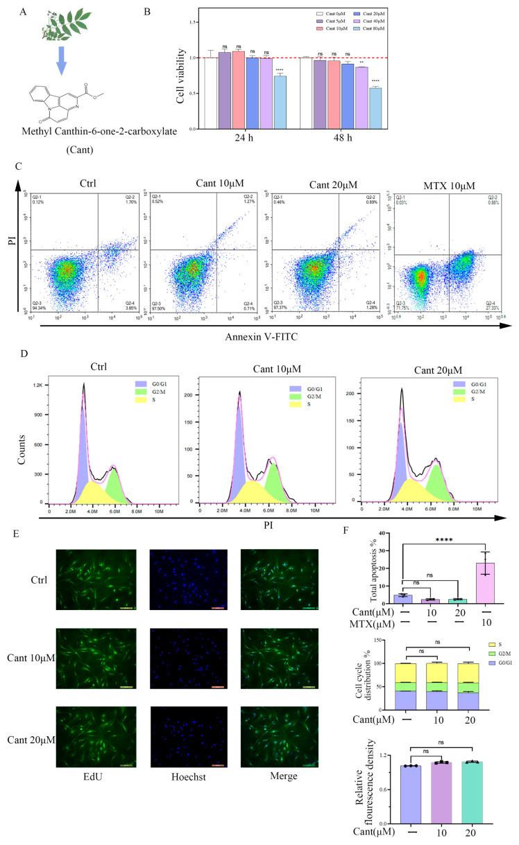
Cant does not increase proliferation, cell cycle, or apoptosis of RA-FLS cells. (A) Chemical formula of canthin-6-one. (B) RA-FLS cells were incubated with various doses of Cant (0, 10, and 20 μM) for 24 h or 48 h. Then, CCK8 assay was performed to examine the impact of Cant on cell proliferation. (C) Annexin V-FITC/PI double staining and flow cytometry were used to determine the status of apoptosis. The result of MTX-treated cells was used as a positive control. (D) Cell cycle was detected by flow cytometry with PI staining. (E) EdU tests were used to assess the status of RA-FLS cell proliferation. Representative photographs of RA-FLS cells stained with EdU (green) and DAPI (blue, ×100) were shown. (F) Quantified data of the effects of Cant on apoptosis, cell cycle, and proliferation of RA-FLS determined by annexin V-FITC/PI, PI, and EdU, respectively. All experiments were performed independently three times. The data are shown as mean ± SEM. ** p < 0.01, **** p < 0.0001, in comparison to RA-FLS without Cant treatment.

