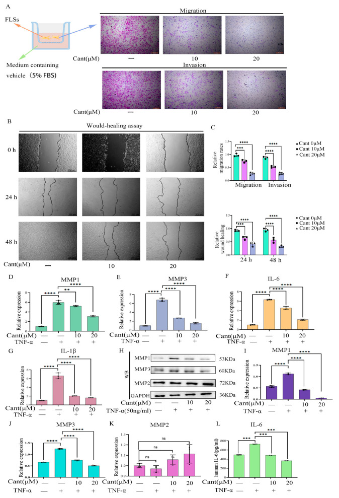
Figure 2.
Cant inhibited migration/invasion, and the production of MMPs and pro-inflammatory cytokines by RA-FLS cells. (A,B) The effects of different concentrations of Cant on RA-FLS cell migration/invasion were determined by transwell (A) and wound-healing assay (B), respectively. The displayed images are representatives from three independent experiments (×100). (C) The migrated or invaded cells in five random fields of each replicate (upper) were quantified, and the statistical chart of the wound-healing results (lower) is shown. The effect of Cant on the transcription of MMP1 (D), MMP3 (E), IL-6 (F), and IL-1β (G) induced by TNF-α (50 ng/mL) by RA-FLSs was determined by qRT-PCR. (H) The levels of MMP1 and MMP3 in Cant-stimulated RA-FLSs were detected by Western blot analysis. The intensity values of the bands of MMP1 (I), MMP3 (J), and MMP2 (K) in (H) were quantified and subjected to statistical analysis. (L) The release of IL-6 in the supernatant of Cant-treated cells was detected by ELISA. All experiments were conducted independently three times. The data are shown as mean ± SEM. *** p < 0.001, **** p < 0.0001, in comparison to RA-FLS cells without TNF-α or Cant treatment (C). ** p < 0.01, *** p < 0.001, **** p < 0.0001, compared with RA-FLS treated with TNF-α alone (D–L).

