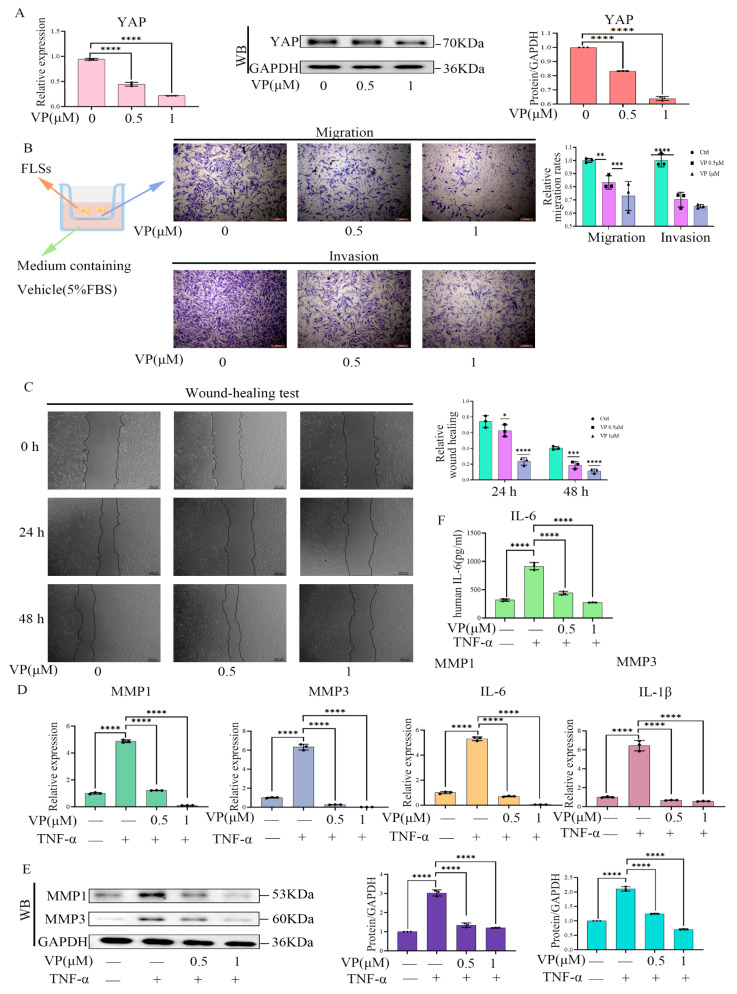
Figure 6.
VP treatment resulted in reduced inflammatory functions of RA-FLS cells. (A) Western blot and qPCR were used to assess the effects of VP (0.5 and 1μM) on YAP expression in RA-FLS cells. (B,C) RA-FLS cells were stimulated with various concentrations of VP. Then, the cells were subjected to transwell assays to detect vertical migration and invasion of the cells (B). Wound-healing tests were performed to detect cell horizontal migration (C). (D,F) VP (0.5 and 1μM) effects on MMP1, MMP3, IL-6, and IL-1β expression in RA-FLSs were assessed using qPCR (D), and IL-6 in the cell culture supernatants was examined using ELISA (F). (E) Western blot analysis was conducted to detect TNF-α-induced (50 ng/mL) MMP1 and MMP3 expression in RA-FLS after VP treatment for 48 h. All experiments were conducted independently three times. The data are shown as mean ± SEM. * p < 0.05, ** p < 0.01, *** p < 0.001, **** p < 0.0001, in comparison to si-NC RA-FLS group (A–C). **** p < 0.0001, in comparison to si-NC group with TNF-α treatment (D–F).

