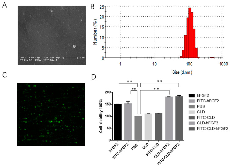
Figure 2.
Microstructural observation and activity analysis of CLD-hFGF2. (A) Ultrastructural observation of CLD-hFGF2 using TEM; (B) analysis of particle size of CLD-hFGF2 with DSL; (C) detection of the protein for CLD-hFGF2 freeze-dried powder with Western blot; and (D) assay of cell viability of CLD-hFGF2 and CLD-hFGF2 freeze-dried powder. (** p < 0.01).

