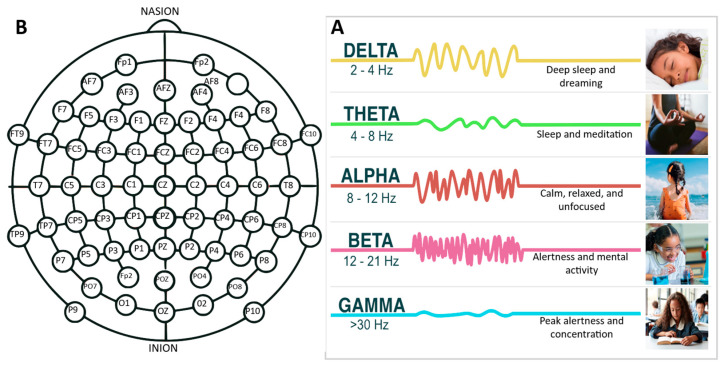
Figure 3.
(A) A detailed schematic representation of electrode placements across the scalp, highlighting standardized locations. This section delineates common configuration points that are essential for ensuring consistent and comparable data across research and clinical studies. (B) An informative chart elucidating various EEG frequency bands—delta, theta, alpha, beta, and gamma—and their associated cognitive and physiological activities. This component serves to emphasize the distinct brain activities and states associated with each frequency range.

