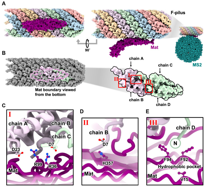
Figure 3.
The interaction of MS2 and the F-pilus [31]. (A) The interaction of MatMS2 (purple) and F-pilus, represented with five different colors for each helical strand. (B) Zoom-in view of panel A focusing on the binding site where MatMS2, shown as a purple boundary, is interacting with four pilin subunits. (C–E) Zoom-in views from panel B, denoted by roman numerals from I to III, rotated 90° to illustrate the reported contacts between MatMS2 and pilin subunits.

