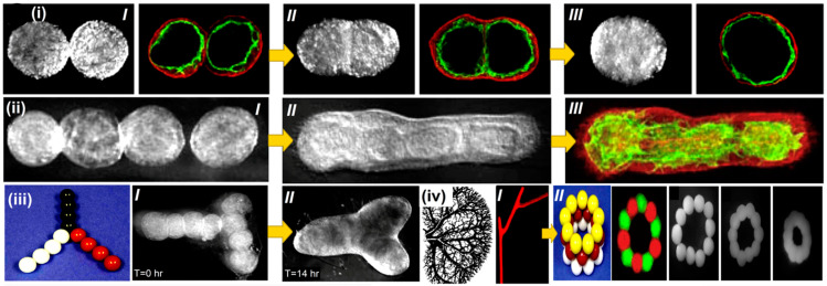
Figure 5.
Bioprinting intraorgan branching vascular trees with uni-lumenal vascular tissue spheroids. Developmental stages of a ring-shaped vascular architecture during tissue fusion. (i) [I–III] Bioengineered vascular tissue spheroids in the shape of rings made from human smooth muscle cells. To show that no cellular mixing occurred during tissue fusion, tissue spheroids were fluorescently stained with green and red fluorescent stains, respectively. (ii) [I] Uni-lumenal vascular tissue spheroids fusing together in a hanging droplet. (ii) [II–III] Successive procedures for vascular tissue spheroids in collagen type 1 hydrogel fusing together. (iii) Physical representation of the development of branched vascular segments from spheroids of uni-lumenal vascular tissue in the production of type 1 collagen hydrogel (iii) [I] initially; and (iii) [II] after the integration of tissues). (iv) Kidney intraorgan vascular tree segment bioprinting employing solid vascular tissue spheroids. [I] A piece of a vascular tree that was bioprinted. [II] A bioassembly model of a tubular vascular tissue construct in 3D employing spheres of solid tissue. Reprinted with permission [113].

