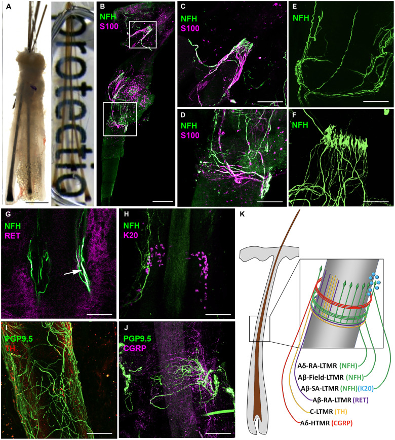
Fig. 1. Human occipital scalp hair follicles are innervated by several LTMRs and an HTMR.
(A) Example of a human follicular unit from scalp skin composed of terminal hairs before (left) and after (right) clearing process. (B) Follicular unit stained with NFH and S100 showing longitudinal (C) and circumferential (D) nerve endings from Aδ-RA-LTMR, Aβ-RA-LTMR, and Aβ-Field-LTMR. (E) Volumetric stacking of human follicular units showing LTMR circumferential endings. (F) Whole-mount images created using volumetric stacking of human follicular units and 3D rendering show the lanceolate shape of LTMR longitudinal endings. (G) Overlap (arrow) of RET and NFH antibody staining reveals Aβ-RA-LTMR longitudinal nerve ending. (H) NFH antibody reveals Aβ-SA-LTMR innervating K20+ Merkel cells. (I) Antibody against TH identifies C-LTMR longitudinal endings (J) Calcitonin gene-related peptide (CGRP) antibody identifies Aδ-HTMR forming circumferential endings around human hair follicles. (K) Schematic representation of the different LTMRs and high-threshold mechanoreceptor (HTMR) that can innervate human hair follicles. While depicted on the schematic for visual clarity, we do not know if these nerves endings are all present on every terminal hair follicle or if there is heterogeneity between follicles in terms of their innervation. Scale bars, (A) 500 μm, (B) 200 μm, (B, i) 100 μm, and [B (ii) and C to H] 50 μm.

