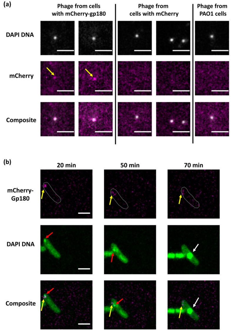
Figure 3.
Localization of PhiKZ vRNAP in infected cells. (a). Co-localization of DAPI-stained DNA and the mCherry signals in PhiKZ phage particles in the lysate of mCherry-gp180-containing cells revealed by fluorescent microscopy. The scale bars are 1 μm. Phage particles from the lysate of mCherry-containing and native PAO1 strain cells are shown as controls. (b). Images of PAO1 cells infected with mCherry-Gp180-containing phiKZ phage. Yellow arrows point to mCherry-Gp180, red arrows point to the area of concentrated DNA, and white arrows to phage nucleus. Times indicate minutes post infection. In the top row, cell contour is delineated in gray. The scale bars are 2 μm.

