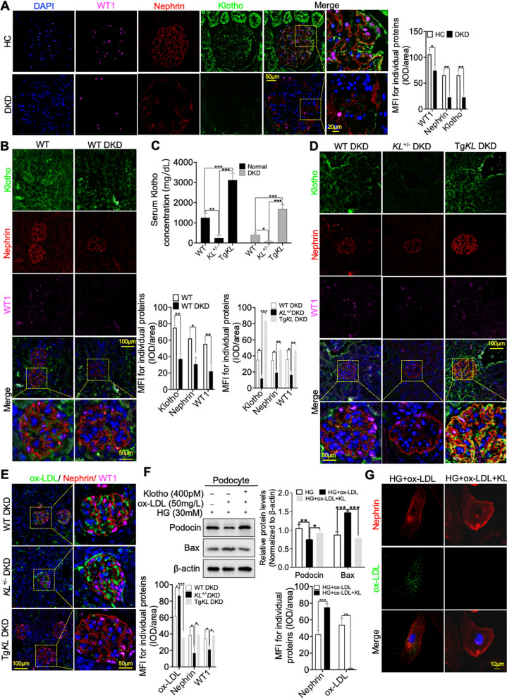
Fig. 2.
Klotho exhibited the potential to alleviate podocyte injury aggravated by renal ox-LDL deposition in DKD. A, B IF staining was conducted using WT1 and Nephrin to evaluate the expression of Klotho on podocytes in the kidneys of DKD patients and mice, in comparison to HC and WT. C Serum Klotho levels in normal and DKD mice were detected by ELISA assay. D Additionally, IF analysis using WT1 and Nephrin was carried out to assess the expression of Klotho in podocytes of WT DKD, KL+/− DKD and TgKL DKD, respectively. E Furthermore, IF analysis using WT1 and Nephrin was performed to evaluate the effect of Klotho on the deposition of ox-LDL in podocytes of the three groups, respectively. F Representative western blot and summarized data showing the effects of Klotho on the relative protein levels of Podocin and apoptosis-associated, Bax in podocytes stimulated with HG and ox-LDL. G Immunofluorescence staining analysis of Klotho highlighting its capacity to mitigate ox-LDL deposition and podocyte injury induced by co-stimulation with HG and ox-LDL. ns no significant; *P < 0.05; **P < 0.01; ***P < 0.001

