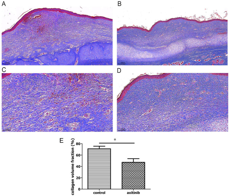
Figure 4.
Histopathological images of Masson stained HS after treatment. (A–D) In contrast with that in the control group ((A), ×40, (C), ×100, n = 20), collagen fibers were loose and regularly arranged in the axitinib group ((B), ×40, (D), ×100, n = 20). (E) The value of collagen volume fraction (CVF, %) was significantly decreased in the axitinib group (n = 20 per group). *P < 0.05.

