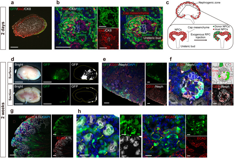
Fig. 2. Successful integration and differentiation of donor cells in the host neonatal nephrogenic niche.
a, b Immunostaining of the chimeric niche in the neonatal kidney 2 days after injection of renal progenitor cells (RPCs) (P3.5). In the outermost layer, the cap mesenchyme consisted of a mixture of host nephron progenitor cells (NPCs, Six2+, GFP −) and donor NPCs (Six2 +, GFP +) surrounding the host ureteric bud (CK8 +, GFP −). c A schematic of (a) and (b). d Fluorescence stereomicroscopic images of the surface and the longitudinal section of host kidney 2 weeks after injection. The yellow dotted lines encircle the host renal cortex. e–h Immunostaining of (d). e, f Chimeric glomeruli containing exogenous podocytes (Neph +, GFP +), nourished by the host endothelial cells (CD31 +, GFP−). g, h Chimeric proximal tubules (LTL +) and distal tubules (ECAD +) indicated by yellow arrows. Scale bars, 500 μm in (a), 50 μm in (b), 2 mm in (d), 100 μm in (e), 10 μm in (f), 200 μm in (g), and 20 μm in (h). CK8, cytokeratin 8; DAPI, 4’,6-diamidino-2-phenylindole; ECAD, E-cadherin; Endo, endothelial cell; GFP, green fluorescent protein; LTL, lotus tetragonolobus lectin; Neph, Nephrin; Podo, podocyte; Six2, sine oculis homeobox homolog 2.

