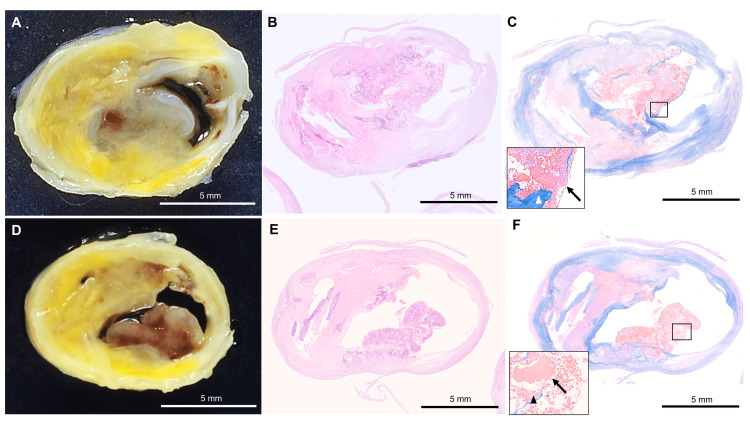
Figure 2. Pathological findings of the calcified nodule in the internal carotid artery.
(A, D) Macroscopically, the internal carotid artery serial cross-sections show an eruptive mass protruding into the lumen with an irregular surface. (B, E) The histopathological finding shows a calcified lesion with nodular calcification protruding into the lumen area on the hematoxylin-eosin (HE) stain. (C, F) The inset shows fibrous cap disruption (triangle) from eruptive calcific nodules associated with a fibrin thrombus (arrow) on Masson's trichrome staining. (B) and (C) are histological images corresponding to (A). (E) and (F) are histological images corresponding to (D). Scale bars: 5 mm.

