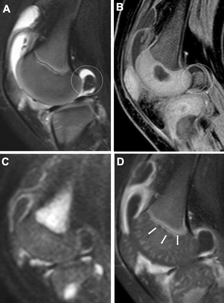
Fig. 8.
MRI for synovitis. A–D. A 1-year-old infant with painful left knee. Fat-suppressed T2-weighted (A) and water selective gradient echo sagittal images (B) showed a large pannus in the posterior fossa (circles, A, B). The articular cartilage was differentiated from the epiphyseal cartilage on both sequences. Diffusion-weighted (C) and Gd-enhanced T1-weighted sagittal images (D) showed high intensities of the synovium, indicative of active synovitis, whereas the popliteal pannus appears black and indistinguishable from joint fluid on these sequences. Vascular structures in the epiphyseal cartilage were well seen on Gd-enhanced image (arrows, D)

