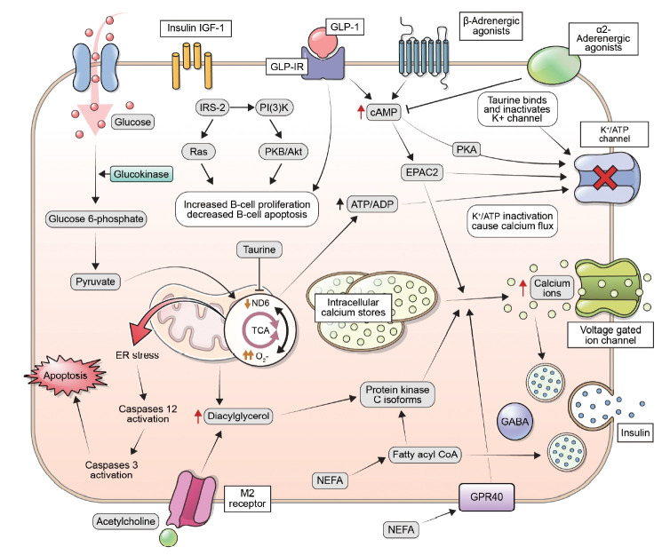
Fig. 1.
Insulin secretion in response to an increase in glucose levels occurs through a process known as glucose-stimulated insulin secretion. This process is mediated by changes in the ratio of adenosine triphosphate (ATP) to adenosine diphosphate (ADP) within the β-cells, which leads to the closure of potassium-ATP channels, depolarization of the plasma membrane, and an increase in cytoplasmic calcium concentrations. These changes ultimately result in the exocytosis of insulin-containing secretory granules. Additionally, in situations where insulin demand is high, β-cells can increase their glucose metabolism through the activation of the enzyme glucokinase. Insulin release is regulated by a variety of mechanisms, including glucose levels, fatty acids, incretins, and nerve signaling. Increased levels of glucose can cause an increase in citrate levels, leading to elevated levels of malonyl-coenzyme A (CoA) and a decrease in carnitine palmitoyl transferase-1 (CPT1) activity. This results in an accumulation of long-chain acyl-CoA and activates protein kinase C (PKC) signaling. Fatty acids can also regulate insulin secretion by signaling through G-protein-coupled receptor 40 (GPR40), or by being metabolized into fatty acyl-CoA and triggering insulin granule exocytosis. The hormone glucagon-like peptide-1 (GLP-1) enhances insulin release in response to glucose via activation of its G protein-coupled receptor. This leads to stimulation of protein kinase A (PKA) and guanine nucleotide exchange factor exchange protein activated by cyclic-AMP (EPAC2). Additionally, acetylcholine release from parasympathetic nerves boosts insulin release through activation of the M2 muscarinic receptor, involving diacyl glycerol (DAG) and PKC. The role of sympathetic nerves in insulin secretion involves changes in adenylyl cyclase and cyclic adenosine monophosphate (cAMP) levels. α2-Adrenergic agonists inhibit insulin secretion, while β-adrenergic agonists stimulate it. Additionally, insulin/insulin-like growth factor 1 (IGF-1) receptor signaling and GLP-1 receptor (GLP-1R) signaling can positively regulate β-cell mass through transactivation of the epidermal growth factor receptor and stimulation of the insulin receptor substrate 2 (IRS-2) pathway. PI3K, phosphoinositide 3-kinase; PKB, protein kinase B; ND6, NADH dehydrogenase subunit 6; TCA, tricarboxylic cycle; ER, endoplasmic reticulum; NEFA, non-esterified fatty acid; GABA, gamma-aminobutyric acid; GPR40, G-protein-coupled receptor 40.

