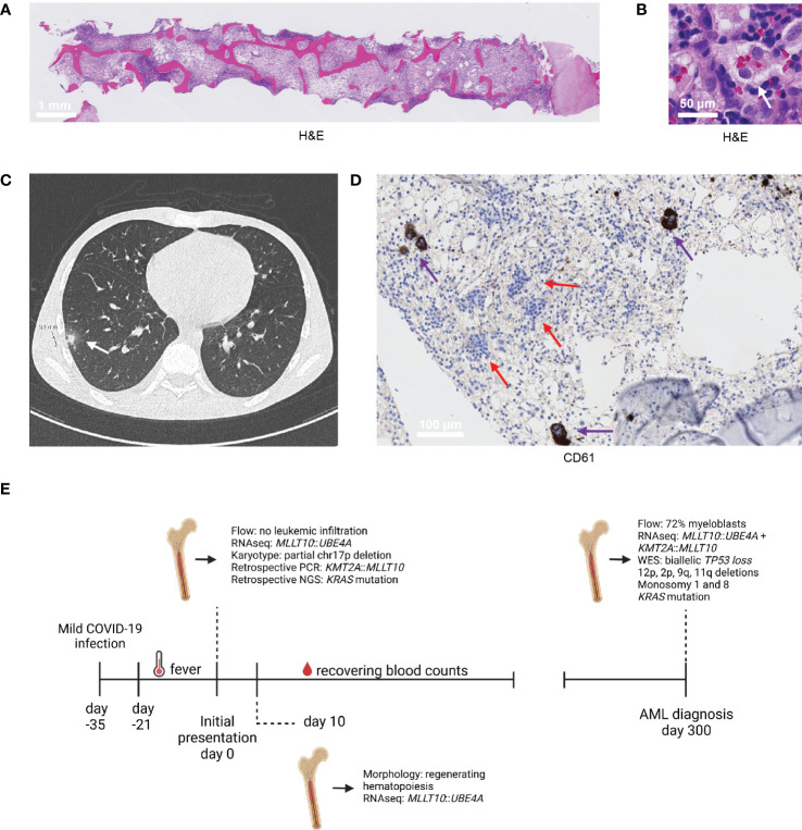
Figure 1.
Diagnostic findings. (A, B) H&E section of the trephine biopsy at initial presentation (A) and an illustration of the hemophagocytosis present in this biopsy, indicated by the white arrow (B). (C) CT scan of the thorax performed at initial presentation, which revealed two nodular lesions in the right lung. One nodular lesion with a diameter of 9 mm is shown (white arrow; the other lesion was similar and is not shown). (D) Immunohistochemistry stain of megakaryocytes (CD61, indicated by the purple arrow) in the trephine biopsy collected at 10 days from initial presentation. The clusters with blue cells, indicated by the red arrows, indicate recovering erythropoiesis. (E) Timeline of the most relevant diagnostic findings.

