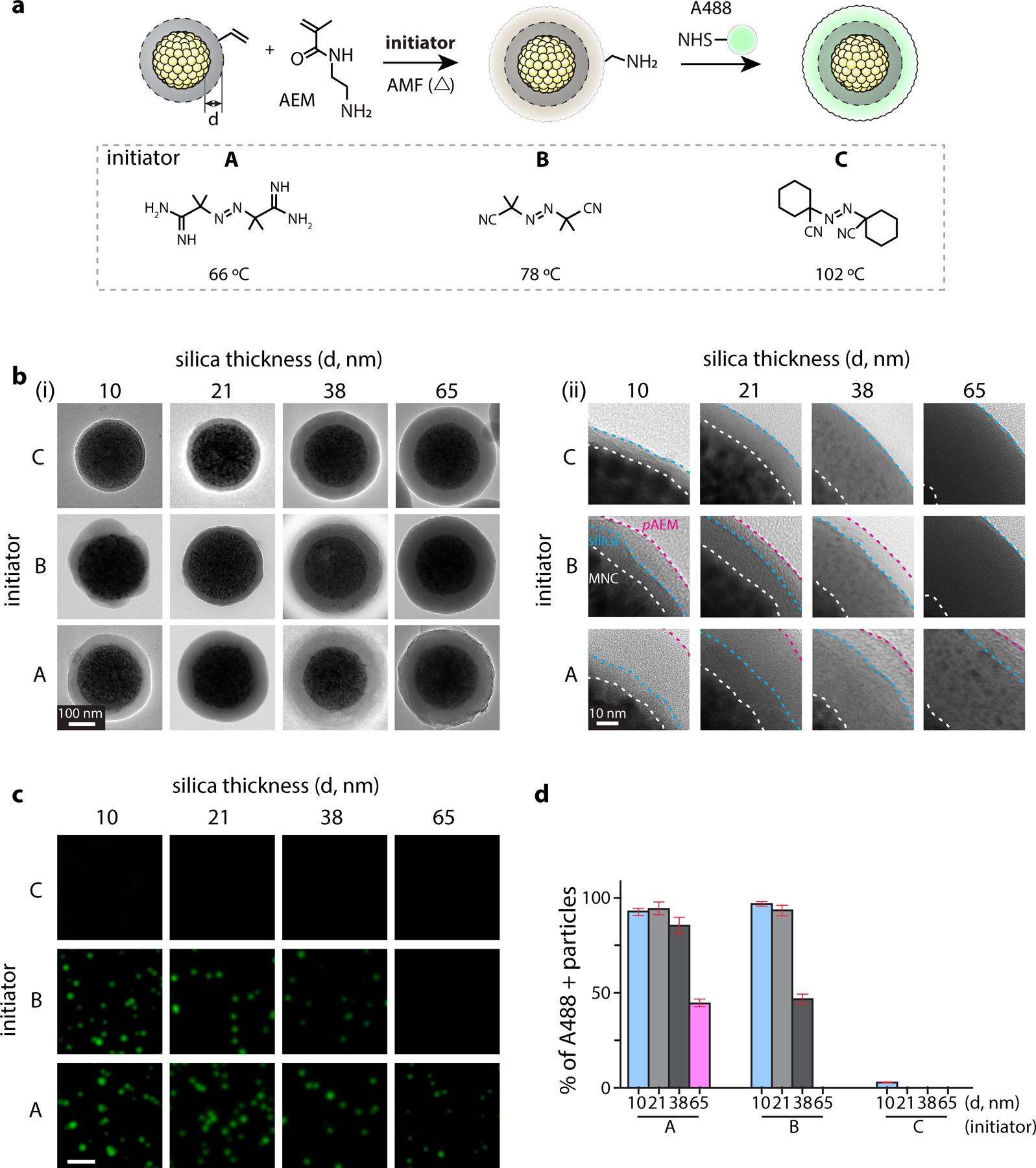
Figure 2. Thermal polymerization of N-(2-aminoethyl)methacrylate (AEM) on MNC@SiO2 to investigate AMF-induced local heating effects by MNCs.

(a) (top) Schematic illustrations of radical polymerization of AEM and subsequent conjugation of amine-reactive fluorescence dyes within it. We varied the SiO2 layer thickness (d = 10, 21, 38, 65 nm) to investigate distance-dependent thermal decay from MNC surface. (bottom) Three thermo-labile azo-molecule initiators with varied decomposition temperatures for AEM polymerization: A, 2,2′-azobis(2-methylpropionamidine) dihydrochloride; B, 2,2′-azobis(2-methylpropionitrile); C, 1,1′-azobis(cyclohexanecarbonitrile). (b) Representative TEM images (Left: entire particle view, Right: zoom-in view) after AMF-stimulation in presence of respective MNC@SiO2 particles and initiators. Scale bar = 100 nm. Interface of MNC/SiO2, SiO2/pAEM, and AEM/vacuum (white dotted line) are marked by white, blue, magenta dotted lines, respectively. Scale bar = 10 nm. (c) Fluorescence signals of the Alexa488-positive particles prepared with radical polymerization conditions shown in Figure 2b and subsequent conjugation of A488–NHS. The images were acquired with a 488-nm laser and FITC emission filter. Scale bar = 2 µm. (d) Fraction analysis of A488-positive particles. Fluorescence-positive particles were identified by using a threshold calculated as mean ± 3×SD of basal signal intensity. Data are mean ± SD from n = 3 independent experiments.
