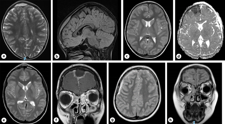
Fig. 1.
Patient 1: axial preoperative T2 (a) and sagittal FLAIR MRI sequences (b), demonstrating midline frontal extradural empyema. Patient 2: axial preoperative T2 MRI sequence, demonstrating right frontal subdural empyema (c) and underlying restricted diffusion on DWI (d). Patient 3: axial preoperative T2 sequence MRI, demonstrating frontal extradural collection, frontal sinusitis, and midline subgaleal extension (e); confirmed on coronal FLAIR (f). Patient 4: axial preoperative T2 sequence MRI highlighting right-sided frontal subdural empyema (g), with evidence of right-sided orbital abscess on preoperative coronal FLAIR (h).

