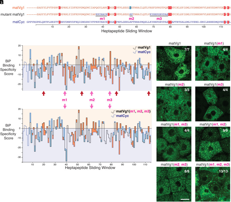
Fig. 4.
Binding motifs for BiP promote ER retention of the Vg1 mature domain. (A) Amino acid sequence alignment of the mature domains of Vg1 (matVg1) and Cyc (matCyc) and mutant matVg1. Cysteines (red) and a potentially glycosylated asparagine (blue) are highlighted. (B and C) Difference charts of the BiP binding specificity scores (34) between two protein sequences along a sliding window of seven amino acids. Orange fills indicate the matVg1 or matVg1(m1, m2, m3) score (black line) > matCyc score (blue line). Conversely, blue fills indicate matCyc score > matVg1 or matVg1(m1, m2, m3) score. Arrows denote regions where matVg1 score > 0 and matCyc score < 0. Red arrows specifically denote regions that contain cysteines involved in cystine-knot formation. (B) Differences in BiP binding specificity scores between matVg1 (black line) and matCyc (blue line). (C) Differences in BiP binding specificity scores between mutant matVg1(m1, m2, m3) (black line) and matCyc (blue line). Note the loss of orange fills in m1, m2, and m3 regions (pink arrows) when compared to (B). (D) Fluorescence images of fixed Mvg1 embryos injected with 50 pg mRNA of sfGFP-tagged vg1 mature domain variants. sfGFP was inserted upstream of the vg1 mature domain in all constructs. (Scale bar, 20 µm.)

