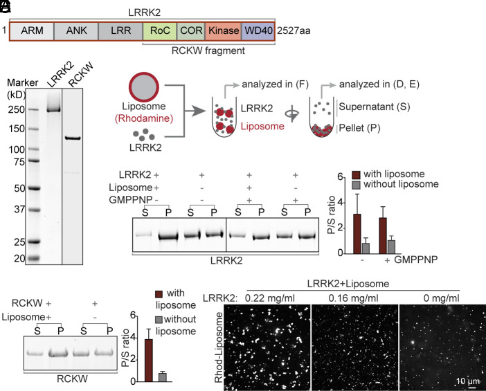
Fig. 1.
Purified LRRK2 binds liposomes. (A) Domain cartoon of full-length human LRRK2. (B) LRRK2 and the RCKW fragment were purified from Expi293 cells and analyzed by SDS-PAGE and CB staining. (C) Schematic diagram of the protein-liposome binding assay. (D and E) Left, Supernatants (S), and pellets (P) of the centrifugation step were analyzed by SDS-PAGE and CB staining. Right, Quantification of the intensities of the gel bands as shown in the Left. Bars represent the ratio between pellets and supernatants (P/S). Values represent mean ± SD n ≥ 3 independent experiments. (F) Confocal microscopy analysis of rhodamine (Rhod)-labeled liposomes incubated with different concentrations of LRRK2, showing LRRK2-induced liposome clusters.

